Short Description
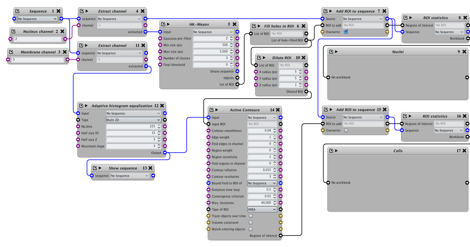
This protocol first extracts the cell nuclei from a given fluorescence channel (full labeling), and grows a contour from each nucleus to extract the cell edge in another fluorescence channel (membrane-labeling).Documentation
Description:
This protocol extract cells in fluorescence images by exploiting 2 types of information:
- fluorescence coming from the entire nucleus
- fluorescence coming from the cell membrane
The procedure is to first extract the nuclei, then grow a contour that will stop when it fits the cell membrane.
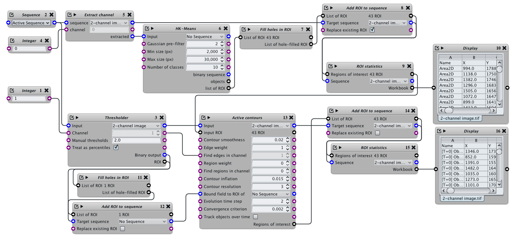
The protocol can be tested on the following image (click on this link to download it).
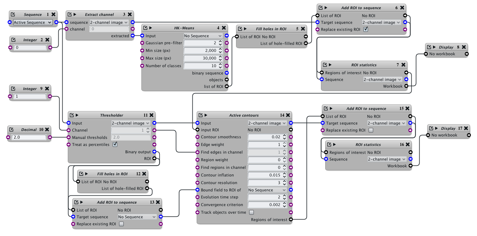
The outcome of the protocol is in blocks 8 and 17: it holds a list of objects and the Intensity statistics over all image channels. This is particularly convenient to compute the expression level of a given marker in one or more extra fluorescence channels.
Important parameters:
- Block 2 (Integer): The nuclear fluorescence channel. Remember, channel indexes start at 0
- Block 4 (HK-Means): The minimum and maximum area (in pixels) of a typical nucleus (depends of your magnification rate)
- Block 9 (Integer): The membrane fluorescence channel. Remember, channel indexes start at 0
- Block 10 (Decimal): The low-pass percentage of intensity used to determine the boundary of the cell layer in the membrane channel
The following parameters are not less important, and are used to adjust the behavior of the growing contours to best match your data, and are all in Block 14 (Active Contours):
- 'Contour smoothness' indicates how smooth the contour should look like, ranges from 0 (no constraint) to 1 (extremely smooth)
- 'Contour inflation' indicates the contour growth factor. 0 is none, positive values will make them grow, negative will shrink them. the value should be kept close to 0, otherwise the contour will not stop when it finds a cell membrane (it's all about balancing this term...)
- 'Evolution time step' indicates how fast the contours should move given the previous parameters (this is an iterative process). Obviously, faster is better, but higher values increase the risk of the contours being somewhat unstable and 'shaking' of even disappearing during the evolution.


